

ThomasS's Store
Owned by ThomasS Joined May 2023 21 Listings
ThomasS's Store
easyid.me/biz/thomass-0742e3 • Verified Store
Scan to open official EasyID digital store, view live catalog, or save business card.
Verified Merchant
EasyID Certified Seller
Fast 24h Packing
Direct courier dispatch
Zero Escrow Direct
Direct merchant settlement
Store Listings (4)
Procollagen Type III (PIIINP)
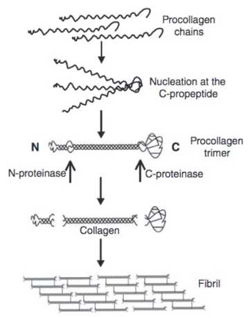
During synthesis of collagen, procollagen undergoes enzymatic cleavage at carboxyl and amino terminal ends by procollagen C-peptidase and procollagen N-peptidase and peptides are released into serum. Procollagen III Peptide is synthesized in fibroblasts as a precursor of collagen type III and then released. <br> <br>In the healthy human liver, the most abundant collagens are the fibril-forming types I and III. Collagen types I and III are synthesized as procollagens with a small amino terminal and a larger carboxy-terminal propeptide. Once secreted into the extracellular space, the propeptides are removed by specific endopeptidases, thus allowing integration of the rigid collagen triple helix into the growing fibril. After this process the three-amino acid procollagen type III N-terminal peptide (PIIINP) is cleaved off procollagen type III forming collagen type III. It is released in serum, and for this reason their serum concentrations have been proposed as a useful marker of collagen type I and III synthesis. This is supported by a diversity of clinical observations demonstrating that high serum levels of these peptides reflect ongoing tissue fibrosis. <br> <br>Currently, the serum measurement of procollagen type III N-terminal peptide by radioimmunoassay has been proposed as a useful test for monitoring disease activity and hepatic fibrosis in liver disease. Concentrations of procollagen type III N-terminal peptide in basal membrane are greater during hepatic fibrosis.
Liver Fibrosis
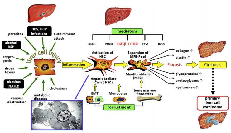
Liver fibrosis is the final common pathway for almost all causes of chronic liver injury. Liver fibrosis is now known to be a dynamic process having significant potential for resolution. Therefore, fibrosis prediction is an essential part of the assessment and management of patients with chronic liver disease. As such, there is strong demand for reliable liver biomarkers that provide insight into disease etiology, diagnosis, therapy, and prognosis in lieu of more invasive approaches such as liver biopsy. <br> <br>Most commonly, fibrosis biomarkers are classified in one of two classes. <br> <br>Class I fibrosis biomarkers are pathophysiological derived from extracellular matrix (ECM) turnover and/or from changes of fibrogenic cell types, in particular, HSCs (Ito-Cells) and their transdifferentiated counterpart. They should reflect the activity of the fibrogenic and/or fibrolytic process and, thus remodeling of ECM. These biomarkers do not indicate the extent of connective tissue deposition, i.e., the stage of fibrotic transition of the organ. Frequently, they are costly laboratory tests and are the result of translation of fibrogenic mechanisms into clinical application. Thus, their selection is driven by hypothesis. <br> <br>Class II fibrosis biomarkers mostly estimate the degree of fibrosis (extent of ECM deposition). In general, they comprise common clinical-chemical tests (e.g., enzymes, proteins, and coagulation factors), which do not necessarily reflect ECM metabolism or fibrogenic cell changes. Their pathobiochemical relation with fibrogenesis is indirect, if at all. Thus, their selection is not driven by hypothesis, but empiric. The markers are standard laboratory tests and are integrated into multiparametric panels. <br> <br>In general, both types of serum biomarkers follow different pathophysiological concepts. Class I markers inform about “what is going on” (i.e., grade of fibrogenic activity); class II markers indicate “where fibrosis is” (i.e., stage of fibrosis).
Acute Inflammation
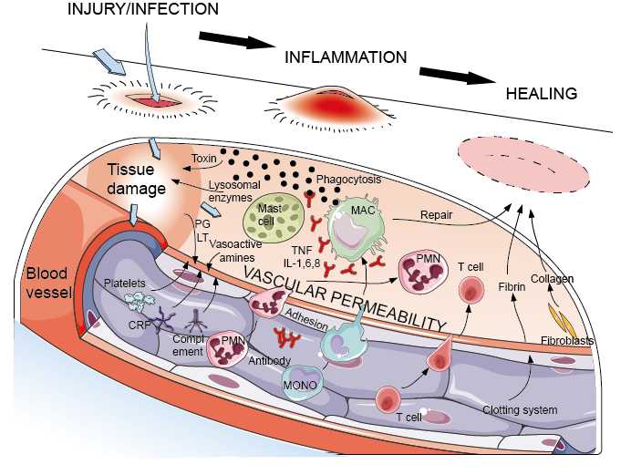
In this simplified scheme, which should be read from left to right, are shown the effects of injury to tissues (Figure 1, top left) and to blood vessels (Figure 1, bottom left). The small black rods represent bacterial infection, a very common cause of inflammation and of course a frequent accompaniment of injury. Note the central role of permeability of the vascular endothelium in allowing access of blood cells and serum components to the tissues, which also accounts for the main symptoms of inflammation – redness, warmth, swelling and pain. <br> <br>It can be seen that the 'adaptive' (or 'immunological') functions of antibody and lymphocytes largely operate to amplify or focus preexisting 'innate' mechanisms; quantitatively, however, they are so important that they frequently make the difference between life and death. <br> <br>Note the central importance of the tissue mast cells and macrophages, and the blood-derived PMNs. Inflammation is usually localized to the area of injury or infection. Occasionally, e.g. in sepsis, uncontrolled inflammation becomes systemic, and causes severe illness, organ failure and ultimately death. Sepsis remains a serious risk after major surgery. If for any reason inflammation does not die down within a matter of days, it may become chronic, and here the macrophage and the T lymphocyte have dominant roles.
Human Factor X ELISA Kit
For quantitative detection of F10 in serum, plasma, tissue homogenates and other biological fluids.
About ThomasS's Store
No description provided.
0.0
Customer Reviews
See what others are saying about ThomasS's Store. We verify purchases to ensure reviews are genuine.
No reviews yet for this store.